Caring for Your Spinal Anatomy in New Jersey

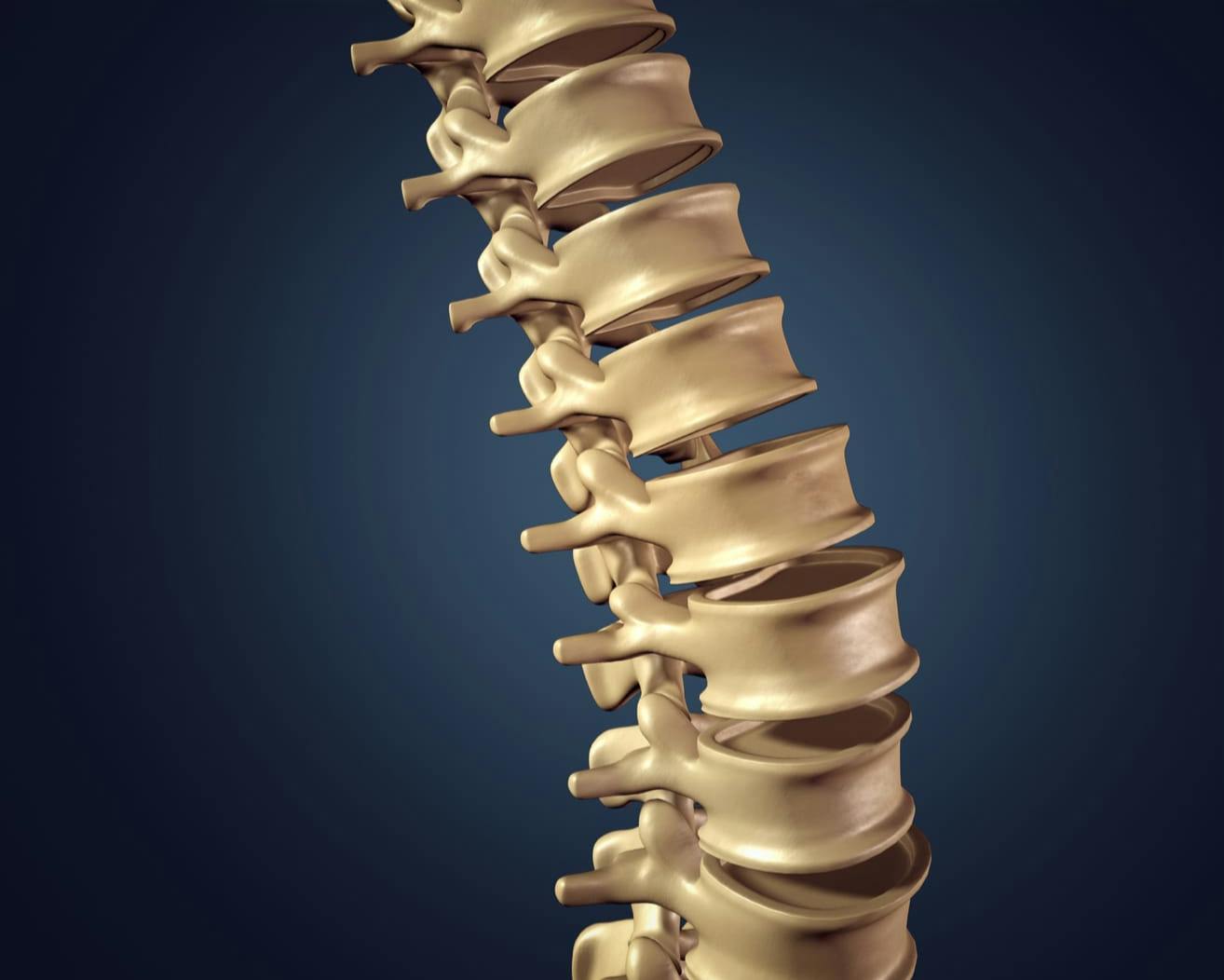
The spine comprises five key regions—cervical, thoracic, lumbar, sacral, and the cauda equina—each serving a distinct purpose. Beneath these broader sections lie intricate networks of nerves, joints, and cushioning discs that work together to keep you upright and mobile. Gaining a clearer view of spinal anatomy will help you better understand your specific condition and guide your decisions about care.

We invite you to contact our Englewood office to schedule a personalized consultation about your spinal health and potential treatment approaches.

Background texture

The Human Spine

While the spine may appear straight from the front, it actually contains subtle curves that help absorb shock and enable a full range of motion. Two primary types of curvature are present:

  • A kyphotic (forward) curve, generally found in the thoracic and sacral regions
  • A lordotic (backward) curve, commonly seen in the cervical and lumbar areas

These curvatures prevent excessive strain on the spine by distributing weight more evenly.

Spinal Column

The spinal column shields critical nerves while supporting the torso and maintaining balance. There are five distinct regions:

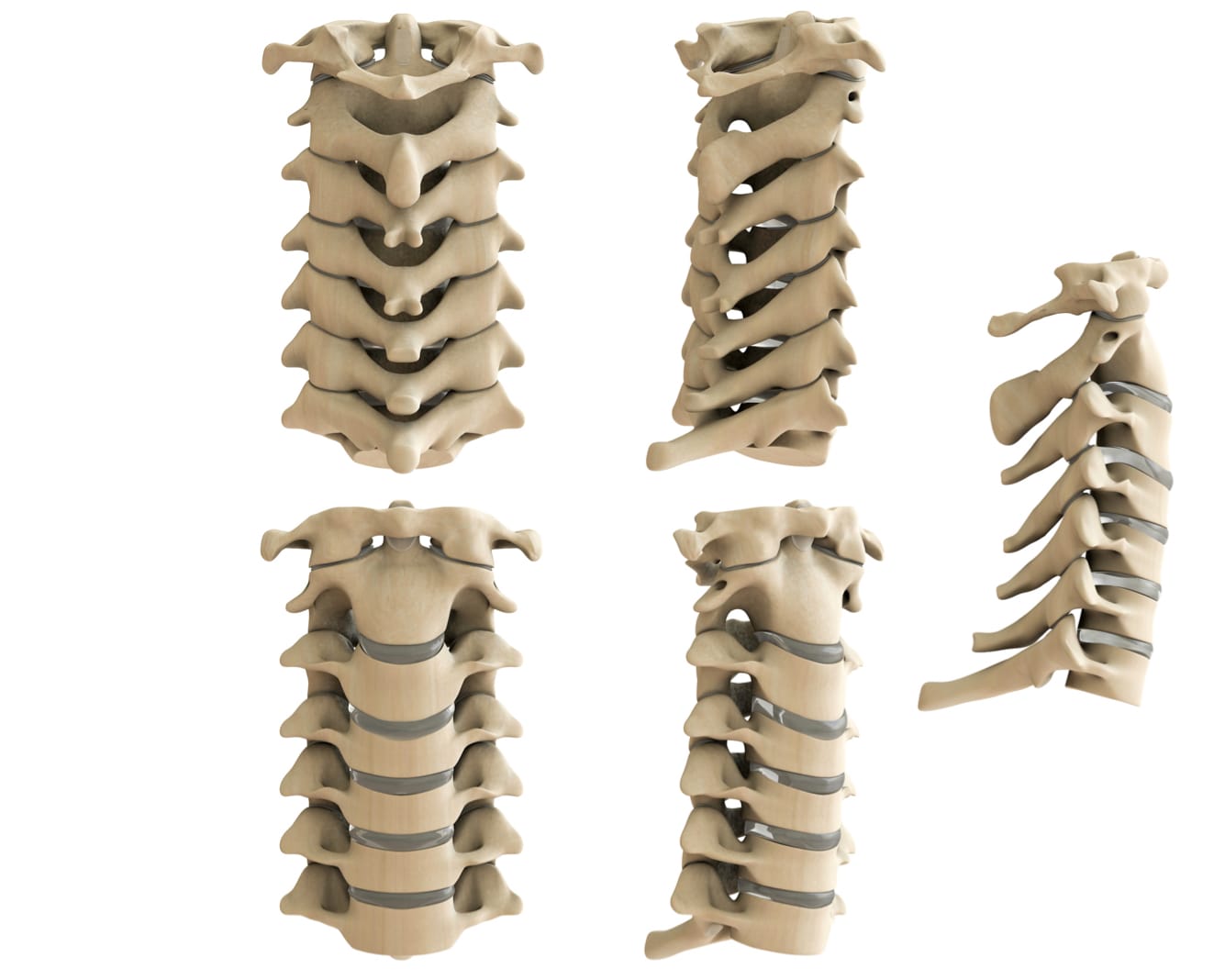
Cervical Spine

Found in the neck, this section supports the skull, enables significant head movement, and protects both the spinal cord and brainstem.

Thoracic Spine

Located in the mid-back and reinforced by the rib cage, this region benefits from extra stability. Though it doesn't rotate extensively, it helps anchor the upper body.

Lumbar Spine

Situated in the lower back, these vertebrae are larger, as they carry much of the body's weight. This zone allows for a wide range of movement—sitting, standing, twisting—but is also more susceptible to strain.

Sacral Spine

Commonly referred to as the tailbone area, the sacrum fuses several vertebrae to provide a stable platform for the spine.

Cauda Equina

Latin for “horse's tail,” this collection of nerve roots lies at the lower end of the spinal cord and supplies sensation and mobility to the legs, bladder, and other structures. When compressed or injured, it may lead to a rare yet serious condition called cauda equina syndrome.

Background media

Spinal Anatomy

Each component of your spine influences posture and movement. Even minor disruptions in vertebrae, discs, or nerves can cause widespread issues in daily life. The spinal cord itself—a cylindrical bundle of nerve tissues—connects with the brain to form the central nervous system. Meanwhile, nerve roots branching outward comprise the peripheral nervous system, delivering signals throughout the body.

Spinal Anatomy

Vertebral Structures

The spine's bones, called vertebrae, have an outer shell of cortical bone (strong and compact) and an inner area of cancellous bone (less dense, honeycomb-like). These vertebrae contain bone marrow that produces blood cells.

Spinal Anatomy

What Are Vertebrae?

Vertebrae are the building blocks of the spine. Each one generally includes:

  • Body: The largest section of the vertebra
  • Pedicles: Two short projections
  • Laminae: Flat plates of bone that help form the back of the vertebral canal

These elements collectively support your weight and allow for coordinated spine movement. Together with other anatomical features—such as processes, facet joints, and discs—the vertebrae help you stand, walk, and remain active.

Processes

Three types of processes attach ligaments and tendons or serve as leverage points for movement:

  • Articular processes connect with neighboring vertebrae.
  • Transverse processes extend out from the sides of the vertebra.
  • Spinous processes project from the back of each vertebra, helping with motion and protection.
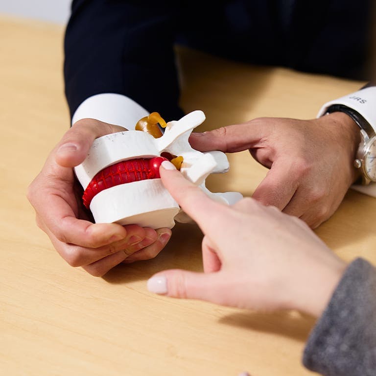
person pointing to a fake spine

Facet Joints, Muscles & Tendons

Facet joints enable bending, twisting, and extending by pairing up the back portions of adjacent vertebrae. Like other joints, each facet joint is wrapped in a connective tissue capsule and produces fluid for smooth movement. In collaboration with intervertebral discs, these joints help regulate flexibility and limit overly extreme motions. Muscles offer stability and power, while tendons anchor muscles to the vertebrae.

Intervertebral Discs, Endplates & Neural Foramen

Intervertebral discs act as shock absorbers between the vertebral bodies, preventing bone-on-bone friction. These discs do not have their own blood supply; they receive nutrients through osmosis. Each disc features:

  • Annulus fibrosus: A durable outer ring
  • Nucleus pulposus: A softer, gel-like center

Endplates on the top and bottom of each vertebra reinforce the intervertebral discs, while neural foramina (openings between vertebrae) offer pathways for nerve roots to exit the spinal column.

Banner media
Doctor media

Why Choose Dr. Stieber for Your Spinal Care?

Dr. Jonathan Stieber has earned a reputation as one of the leading spinal surgeons, recognized for pioneering minimally invasive solutions, motion preservation surgery, and robotic-assisted techniques. With experience as a clinical professor and numerous publications to his name, he delivers advanced, patient-focused care aimed at restoring function and comfort.


We encourage you to reach out to our Englewood team today for a thorough evaluation and a personalized plan designed to support and improve your spinal health.

Schedule a Consultation And Get The Pain-Free Lifestyle You Deserve

NYC spine surgery specialist, Dr. Jonathan Stieber, provides his patients with the most effective treatment options available, all of which are tailored to meet your individual needs. With Dr. Stieber guiding your care, it is possible to say goodbye to pain and enjoy an active lifestyle once again.

Contact Us
Contact Us Page
Accessibility: If you are vision-impaired or have some other impairment covered by the Americans with Disabilities Act or a similar law, and you wish to discuss potential accommodations related to using this website, please contact our Accessibility Manager at (646) 916-3962.
Contact Us