(646) 916-3962 Menu

Pre-Treatment Images

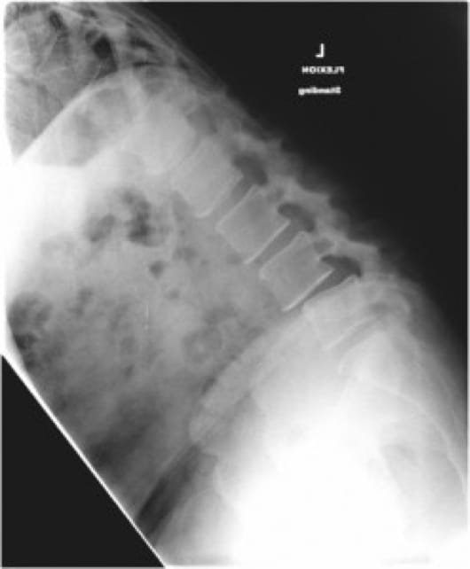
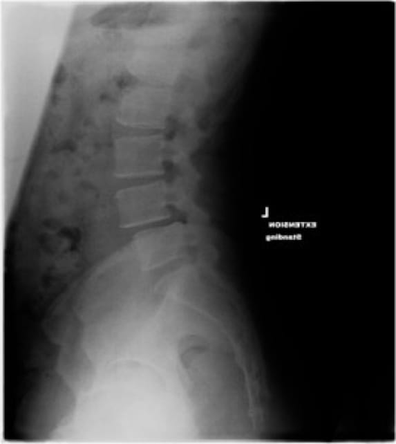
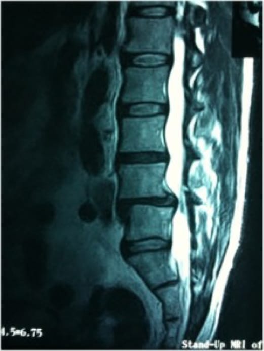
Pre-operative x-rays demonstrated a Grade I degenerative spondylolisthesis with instability on flexion-extension radiographs (Figures 1, 2). MRI findings (Figure 3) revealed disc bulging and uncovering of the intervertebral disc in addition to spondylolisthesis.

Grade 1 Spondylolisthesis, OrthoManhattan

Figure 1

Grade 1 Spondylolisthesis, OrthoManhattan

Figure 2

Figure 1. Standing flexion x-ray from the side (lateral); bending forward at the waist

Figure 2. Standing extension x-ray from the side (lateral); bending backward

Grade 1 Spondylolisthesis, OrthoManhattan

Figure 3

Grade 1 Spondylolisthesis, OrthoManhattan

Figure 4

Figure 3. Lateral MRI demonstrates spondylolisthesis at L4-L5

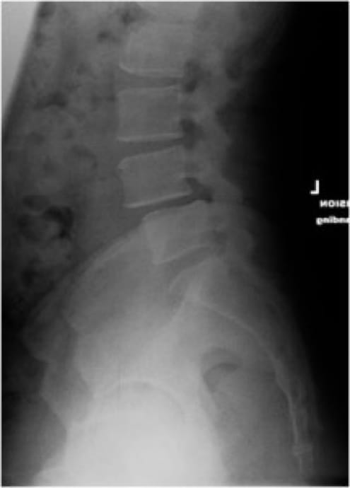
Figure 4. Lateral x-ray

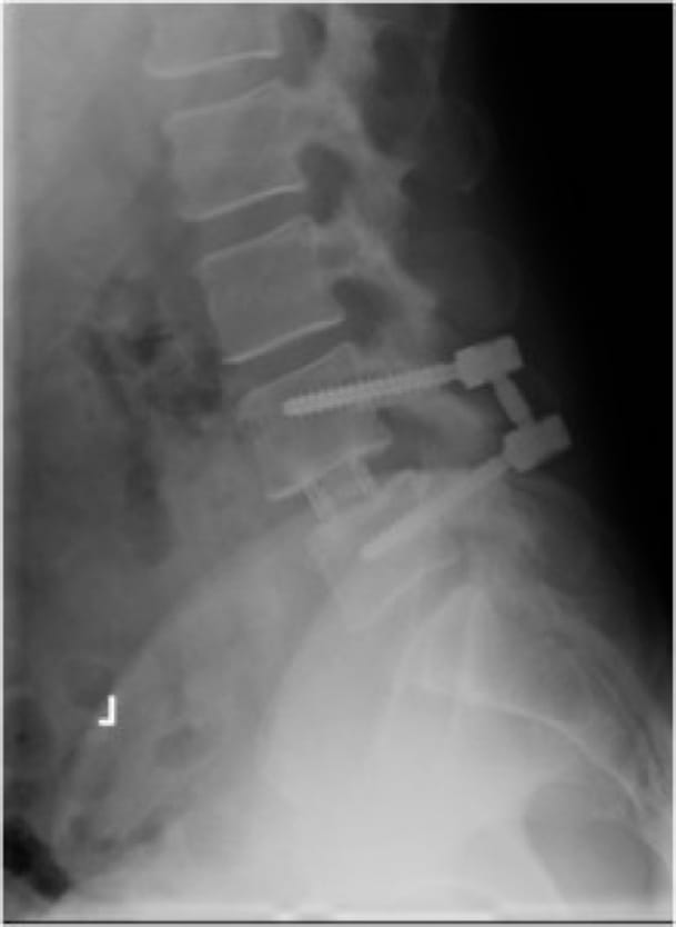
Post-Operative Image

Grade 1 Spondylolisthesis, OrthoManhattan

Figure 5. Post-operative lateral x-ray at 6 months after spondylolisthesis spine surgery. The interbody device can be seen between the fourth and fifth lumbar vertebral bodies (L4-L5)

© Stieber MD. All Rights Reserved. Designed & Developed by Studio III
Alternate Phone: (212) 883-8868

Schedule a Consultation