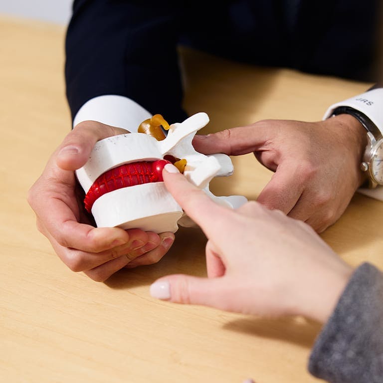
(646) 916-3962 Menu

Understanding Your Spine

There are five main regions of the spine, consisting of the cervical, thoracic, lumbar, sacral, and cauda equina. Each distinct region has its own unique make-up. But what about the spinal nerves, joints, and discs that keep you standing upright? Understanding spinal anatomy will help you understand your specific condition, and how to best seek treatment.


The Human Spine

Even though spines look like they are straight and rigid when viewed from the front, a healthy spine is actually slightly s-shaped, with distinct curves. The curves are necessary for shock absorption and normal movement.

There are two kinds of spinal curves:

  • A kyphotic curve bends forward (normally found in thoracic and sacral spine)
  • A lordotic curve bends backward (normally found in the cervical and lumbar regions of the spine)

Spinal column

One of the most important pieces of physical architecture in the body, the spinal column protects the spine. In addition to protecting the spinal cord and nerve roots, the spinal column provides structural support and balance to maintain an upright posture and enables flexible motion. There are 5 different distinct regions of the spine:

Spinal Anatomy

Cervical Spine

Cervical spine is found in the neck; it supports your skull, protects your brain stem and spinal cord, and helps facilitate a wide range of head movement.

Thoracic Spine

Thoracic spine is located in the chest area. This area of the spine is supported by the strength of the ribcage and does not rotate.

Rib attachments add to the strength and stability of the thoracic spine. The thoracic spine is often less prone to injury because it is braced by the rib cage. Additionally, thoracic vertebrae do not rotate as much as vertebrae in the neck or lower back.

Lumbar Spine

Lumbar spine is your lower back. The vertebrae in this part of your spine are the largest because they bear the most of your body’s weight. This area has a wide range of motion and is responsible for most movement in the body—sitting, standing, pushing, etc.—making it more vulnerable to injury.

Sacral Spine

Sacral spine is found in the sacrum, more commonly referred to as the tailbone.

Cauda Equina

Cauda equina is a collection of nerve roots at the very end of the spinal cord, also known as “horse’s tail.” These nerves are responsible for motor and sensory functions in the bladder and legs.

Although the cauda equina is technically a collection of nerve roots and not vertebrae, if there is trauma or complications with a lumbar herniated disc, cauda equina syndrome (CES) can occur. This is a rare occurrence, but patients with back pain should be aware of the following symptoms:

Although rare, cauda equina syndrome (CES) usually occurs as a result of trauma or complications related to lumbar herniated disc. Patients with back pain should be aware of the following symptoms:

  • Severe low back pain
  • Weakness and/or loss of sensation in the legs or groin
  • Urinary retention; urinary or bowel incontinence

If left untreated, the damage of CES can be severe.

Spinal Anatomy

When one piece of your spinal anatomy isn’t working the way it’s supposed to, your whole body can be affected. This means that, in addition to understanding the different regions of the spine, it’s important to understand how each part of your spine, from your vertebrae to each nerve ending, work together to keep your spine healthy, and your body moving.

The spinal cord is a slender cylindrical structure about the diameter of the little finger, contained and protected within the spinal canal. Your brain and your spinal cord make up your central nervous system, while the nerve roots that branch out of the spine into the body form the Peripheral Nervous System.

Spinal Anatomy

Vertebral Structures

The spine is made up of bones called vertebrae. The outer shell of a vertebra is made of cortical bone, which is solid and strong. Inside each vertebra is cancellous bone, which is less dense and consists of loosely knit structures that resemble honeycomb. Bone marrow, responsible for forming red blood cells and some types of white blood cells, is found inside of cancellous bone.

Spinal Anatomy

What Are Vertebrae?

Vertebrae are the series of small bones that make up the spine. Each vertebra consists of the following common elements:

  • Body: largest part of a vertebra
  • Pedicles: two short processes
  • Laminae: two relatively flat plates of bone

Vertebrae work within the spinal column to keep your body upright and moving naturally. In order to do so, they are supported by the spinal column and several other unique elements of spinal anatomy, including processes, facet joints, intervertebral discs, endplates, foramen, and muscles and tendons.

Processes

Processes assist in the movement of your spine, and there are three types:

  • Articular
  • Transverse
  • Spinous

Articular and transverse processes connect ligaments and tendons. Spinous processes act as a lever to help with vertebral motion.

Facet Joints, Muscles & Tendons

Facet joints help the spine to bend, twist, and extend in different directions. They also help restrict excessive movement, to prevent hyperextension or hyperflexion (e.g., whiplash). Like other joints in the body, each facet joint in the spine is surrounded by a capsule of connective tissue and produces fluid that nourishes and lubricates the joint.

  • Each vertebra contains two facet joints.
  • The surfaces of each facet joint are coated with cartilage that helps them to move smoothly.
  • The facet joints, along with the intervertebral discs, allow for motion in the spine.

Muscles in the vertebral column provide spinal support and stability and serve to flex, rotate, or extend the spine. Tendons, which are similar to ligaments, attach muscles to the bones of the spine.

Intervertebral Discs, Endplates & Neural Foramen

Between each vertebral body is a cushion; the intervertebral disc. Discs absorb stresses the body incurs during movement and prevents vertebrae from grinding against one another (which causes pain).

  • The intervertebral discs are the largest structures in the body that do not have a vascular system.
  • Instead, intervertebral discs receive nutrients through osmosis.
  • Each disc is made up of two parts: the annulus fibrosus and the nucleus pulposus.

The top and bottom of each vertebra is coated with an endplate, which blend into the intervertebral disc and help support it. Neural foramina are hollow passageways between each vertebra, that act as windows for nerve roots to exit the spinal column.

Why Choose Dr. Stieber for your Spinal Care?

Dr. Jonathan Stieber is one of the best spinal surgeons in NYC. He serves as Assistant Clinical Professor of Orthopedic Surgery at The Icahn School of Medicine at Mount Sinai and as Clinical Assistant Professor of Orthopedic Surgery at the NYU School of Medicine.

Skilled in the most current surgical techniques and emerging technologies, Dr. Stieber is globally recognized as a leading orthopedic specialist. He is specifically interested in working with patients to create minimally invasive solutions for the treatment of spinal disorders and motion preservation surgery and is also experienced with robotic spinal surgery. Additionally, Dr. Stieber has published in numerous peer-reviewed papers and presented his research both nationally and internationally.

Spinal Anatomy

© Stieber MD. All Rights Reserved. Designed & Developed by Studio III
Alternate Phone: (212) 883-8868

Schedule a Consultation