

Schedule a Consultation And Get The Pain-Free Lifestyle You Deserve
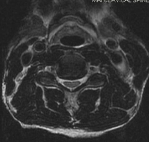
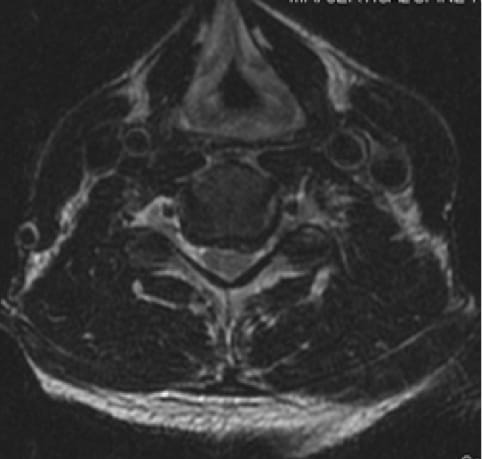
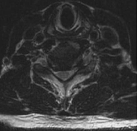
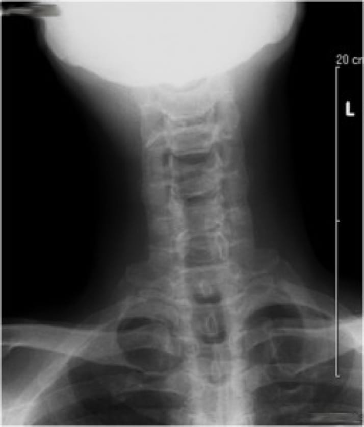
NYC spine surgery specialist, Dr. Jonathan Stieber, provides his patients with the most effective treatment options available, all of which are tailored to meet your individual needs. With Dr. Stieber guiding your care, it is possible to say goodbye to pain and enjoy an active lifestyle once again.
Contact Us